Horner Syndrome Mri Protocol
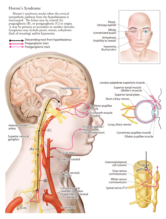
Horner syndrome mri protocol. The present study aims to provide detailed observations on the cavernous segment of the abducens nerve AN emphasizing anatomical variations and the relationships between the nerve and the internal carotid plexus. Physiopedia is not a substitute for professional advice or expert medical services from a qualified healthcare provider. Six-minute magnetic resonance imaging protocol for evaluation of acute ischemic stroke.
An additional five specimens were subjected to histological. MRA found to be superior to MRI with sensitivity of 95 and specificity of 99 overall. A shunt is an abnormal communication between the right and left sides of the heart or between.
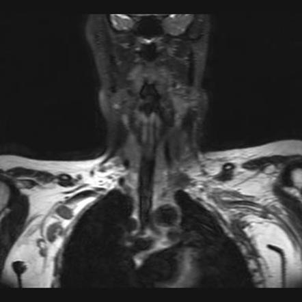
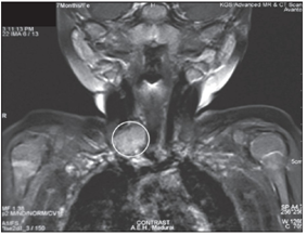
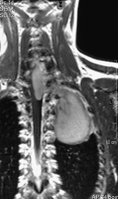
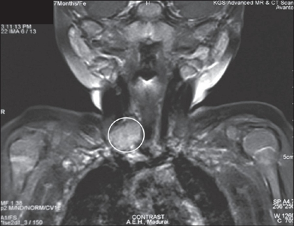
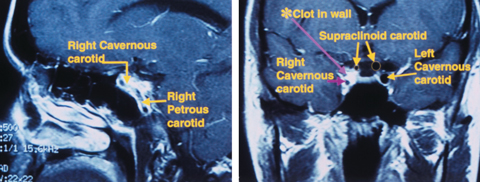
Our follow-up protocol establishes a physical exam every 3 months with basic blood work MRI of the neck and chest every six months to minimize. Atlas of Emergency Radiology Block Jordanov Stack. Cranial nerve palsies in either circulation or a Horner syndrome can be due to ischemia or stretching or compression of nerves from an intramural hematoma.
The content on or accessible through Physiopedia is for informational purposes only. G902 Horners syndrome G904 G909 Opens in a new window Autonomic dysreflexia Disorder of the autonomic nervous system unspecified G950 G9519 Opens in a new window Syringomyelia and syringobulbia Other vascular myelopathies. Scientific Evidence 3e PDF Macintyre PE Schug SA Scott DA Visser EJ Walker SM.
X The modern era of surgical stabilization of rib fractures SSRF was ushered in by the publication of the inaugural randomized controlled trial RCT in 2002 1. Five specimens of the AN were stained using Sihlers method. Since that time interest in SSRF has grown dramatically as evidenced by an over eight-fold increase in use of the operation for patients with a diagnosis of flail chest from 2007 to 2014 2-4.
The brachial plexus is a network of intertwined nerves that control movement and sensation in the arm and hand. The most prevalent opinion is. This process is driven by elevated leptin levels and upregulation of a HIF1α-VEGF signaling axis in local astrocytes.
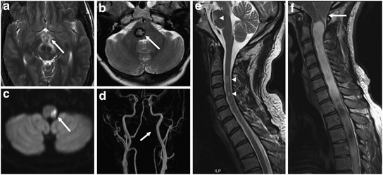
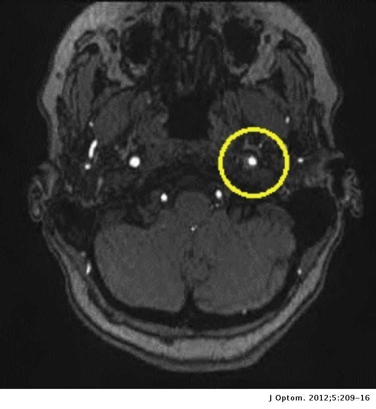
For vertebral injuries specifically MRA was only 60 sensitive. Vertebral artery dissection VAD is a flap-like tear of the inner lining of the vertebral artery which is located in the neck and supplies blood to the brainAfter the tear blood enters the arterial wall and forms a blood clot thickening the artery wall and often impeding blood flowThe symptoms of vertebral artery dissection include head and neck pain and intermittent or permanent stroke.
The creatures their toxins and care of the poisoned patient 2e.
Vertebral artery dissection VAD is a flap-like tear of the inner lining of the vertebral artery which is located in the neck and supplies blood to the brainAfter the tear blood enters the arterial wall and forms a blood clot thickening the artery wall and often impeding blood flowThe symptoms of vertebral artery dissection include head and neck pain and intermittent or permanent stroke. Show that during diet-induced obesity in mice there is a profound remodeling of the gliovascular interface in the hypothalamus resulting in arterial hypertension. Scientific Evidence 3e PDF Macintyre PE Schug SA Scott DA Visser EJ Walker SM. The present study aims to provide detailed observations on the cavernous segment of the abducens nerve AN emphasizing anatomical variations and the relationships between the nerve and the internal carotid plexus. An additional five specimens were subjected to histological. A follow-up and post-treatment monitoring protocol needs to be well outlined due to the 30 rate of local recurrence 8. Prospective protocol in which MRI versus MRA was evaluated in angiographically confirmed BCVI in 18 patients. This process is driven by elevated leptin levels and upregulation of a HIF1α-VEGF signaling axis in local astrocytes. 49 Likes 2 Comments - College of Medicine Science mayocliniccollege on Instagram.
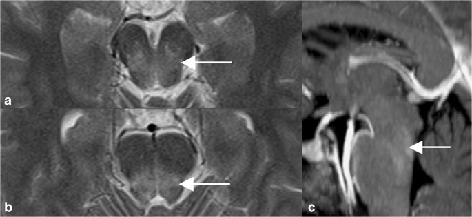
You cant see it but theyre smiling from ear to ear behind those masks. CT angiography and dissection protocol MRI and MR angiography. What the Anesthesiologist Should Know before the Operative Procedure What are right-to-left shunts. Five specimens of the AN were stained using Sihlers method. An additional five specimens were subjected to histological. Physiopedia is not a substitute for professional advice or expert medical services from a qualified healthcare provider. The content on or accessible through Physiopedia is for informational purposes only.














Posting Komentar untuk "Horner Syndrome Mri Protocol"